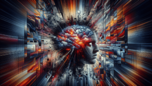
You’ve probably experienced the throbbing pain, nausea, and sensitivity to light that come with a migraine. But have you ever wondered if these debilitating headaches actually show up on an MRI scan? In this article, we will explore whether migraines can be detected through the use of an MRI and delve into the fascinating world of neuroimaging to shed light on this common question. So, sit back, relax, and get ready to uncover the mystery behind migraines and MRI scans.
Understanding Migraines
Migraines are a type of headache disorder that can cause intense pain and a wide range of other symptoms. They affect millions of people worldwide and can significantly impact their quality of life. To better understand migraines, it is important to explore their definition, causes, and common symptoms.
Definition of migraines
Migraines are characterized by moderate to severe headaches that are often accompanied by other symptoms. These headaches typically occur on one side of the head and can last for hours or even days. In addition to the headache itself, migraines may also cause nausea, vomiting, sensitivity to light and sound, and visual disturbances.
Causes of migraines
The exact cause of migraines is still not fully understood. However, researchers believe that a combination of genetic and environmental factors plays a role in their development. Some common triggers for migraines include stress, hormonal changes, certain foods and drinks, changes in sleep patterns, and environmental factors such as bright lights or strong smells.
Common symptoms of migraines
In addition to the severe headaches, migraines can present with a variety of other symptoms. These can include:
- Aura: Some individuals may experience sensory disturbances known as auras before the onset of a migraine. These can manifest as visual changes, such as seeing flashing lights or zigzag patterns, or as unusual sensations in other parts of the body.
- Nausea and vomiting: Many migraine sufferers experience gastrointestinal symptoms such as nausea and vomiting during an attack.
- Sensitivity to stimuli: Migraines can make individuals more sensitive to light, sound, and even certain smells. This sensitivity can exacerbate the pain and discomfort associated with the headaches.
- Fatigue and mood changes: Migraines can cause exhaustion and fatigue, as well as mood changes such as irritability or depression.
The Role of MRI in Diagnosing Migraines
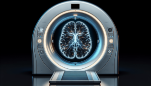
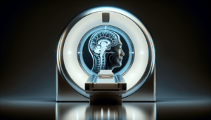
Magnetic Resonance Imaging (MRI) is a powerful diagnostic tool that uses a magnetic field and radio waves to create detailed images of the body’s internal structures. It has become an essential tool in the evaluation and diagnosis of various medical conditions, including migraines. Let’s explore the overview of MRI as a diagnostic tool, its ability to detect migraines, and the benefits of using MRI for migraine diagnosis.
Overview of MRI as a diagnostic tool
MRI is a non-invasive imaging technique that provides a high level of detail and can capture images from different angles. It allows healthcare providers to visualize the structures and tissues within the body, including the brain and its blood vessels. This ability to render detailed images makes MRI an indispensable tool in diagnosing various neurological conditions, including migraines.
MRI scans and their ability to detect migraines
When it comes to migraines, MRI scans can play a crucial role in ruling out other possible causes of headache symptoms and identifying any underlying structural abnormalities or conditions. While migraines themselves may not always be visible on an MRI scan, the images obtained can help provide valuable information to healthcare providers and aid in the diagnostic process.
Benefits of using MRI for migraine diagnosis
MRI offers several benefits when it comes to diagnosing migraines. Firstly, it is a non-invasive procedure that does not use ionizing radiation, making it a safe imaging option for patients. Additionally, MRI can provide highly detailed images of the brain, allowing healthcare providers to evaluate various structures and potential abnormalities. This level of detail can aid in accurate diagnosis, treatment planning, and monitoring the effectiveness of treatments for migraines.
What Can Be Seen on an MRI for Migraines?
MRI scans for migraines can reveal important information about the brain’s structures, blood flow, and the presence of any lesions or tumors. Understanding what can be seen on an MRI can help healthcare providers gain valuable insights into the underlying causes and mechanisms of migraines.
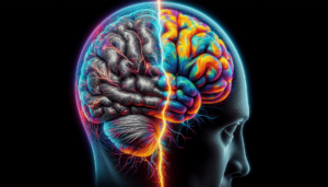
Brain structure and abnormalities
MRI scans can provide detailed images of the brain’s anatomy, allowing healthcare providers to assess its structure for any abnormalities. This can include identifying any structural variations, such as malformations or developmental issues, that may contribute to migraines. Additionally, MRI can help identify any potential sources of pressure on the brain, such as tumors or cysts, which could be causing migraine symptoms.
Blood flow and circulation variations
MRI scans can also assess blood flow and circulation in the brain, providing valuable information about potential vascular causes of migraines. Changes in blood flow patterns, such as arterial spasms or abnormalities in blood vessels, can be detected using specialized MRI techniques. This information can assist in understanding the role of blood flow alterations in triggering migraines and guide treatment approaches.
Presence of lesions or tumors
MRI scans are highly effective in detecting the presence of lesions or tumors in the brain. While these are not common causes of migraines, they can sometimes contribute to headache symptoms. MRI can help healthcare providers identify and characterize these abnormalities, allowing for appropriate treatment planning and management.
Indications for MRI in Migraine Patients
Determining when an MRI is necessary for migraine patients depends on several factors. Healthcare providers consider the indications for ordering an MRI, such as the frequency and severity of migraines, as well as the presence of any neurological symptoms. Let’s delve into the key indications for MRI in migraine patients.
Indications for ordering an MRI for migraines
While not all migraine patients require an MRI, there are certain situations where it may be warranted. These indications include:
- New or sudden-onset migraines: If a patient experiences a sudden change in the frequency, severity, or type of migraines, an MRI may be recommended to rule out any underlying causes or conditions.
- Neurological symptoms: If a patient presents with neurological symptoms, such as weakness, numbness, or changes in vision, an MRI may be ordered to assess the brain for potential causes.
- Atypical symptoms: Migraines with atypical features, such as a prolonged duration or an unusual pattern of symptoms, may prompt healthcare providers to order an MRI to explore other potential causes.
Frequency and severity of migraines
The frequency and severity of migraines can also play a role in determining the need for an MRI. If a patient experiences frequent migraines that significantly impact their daily life, an MRI may be recommended to gain a better understanding of any possible contributing factors. Additionally, if migraines are associated with severe symptoms or complications, an MRI can help identify any underlying issues that require further investigation.
Presence of neurological symptoms
The presence of neurological symptoms, such as weakness, numbness, or difficulty speaking, can be indicators of more serious underlying conditions. When these symptoms accompany migraines, healthcare providers may order an MRI to assess the brain for any structural abnormalities or other potential causes.
MRI vs. Other Diagnostic Techniques
While MRI is a powerful tool in diagnosing migraines, it is important to consider its advantages and disadvantages compared to other diagnostic techniques. Comparing MRI with techniques such as CT scans can help healthcare providers determine the most appropriate imaging approach for each individual case.
Comparison with CT scan
CT (computed tomography) scans and MRI are both imaging techniques that provide valuable information about the body’s internal structures. However, they differ in their mechanisms and image quality. While CT scans use X-rays to create detailed cross-sectional images of the body, MRI uses a magnetic field and radio waves. In terms of migraines, MRI is generally preferred over CT scans due to its superior ability to visualize soft tissues, including the brain, with greater detail.
Advantages and disadvantages of MRI over other techniques
One of the significant advantages of MRI over other imaging techniques is its ability to provide highly detailed images without using ionizing radiation. This makes it a safer option, particularly for individuals who require frequent or repeated imaging. MRI is also better suited for evaluating soft tissues and can provide multi-planar imaging, allowing for a comprehensive evaluation of the brain’s structures.
However, there are some limitations to consider. MRI scans can be more time-consuming than other imaging techniques, and patients need to remain still for an extended period during the procedure. Additionally, some individuals with certain medical implants or metallic devices may not be eligible for MRI due to safety concerns.
When other imaging studies might be preferred
While MRI is generally the preferred imaging modality for evaluating migraines, there are specific scenarios where other imaging studies may be preferred. For example, CT scans may be more appropriate in emergency situations where time is of the essence, as they can provide faster results. Additionally, in cases where there are contraindications to MRI, such as patients with certain metallic implants or claustrophobia, alternative imaging options like CT scans may be considered.
Limitations of MRI in Detecting Migraines
While MRI is a valuable diagnostic tool, it does have limitations when it comes to detecting migraines. Understanding these limitations is essential in interpreting MRI results accurately and avoiding potential false negatives or false positives.
Migraine triggers that may not be visible on MRI
Migraines can be triggered by various factors, including stress, hormonal changes, and certain foods. However, these triggers may not be visible on an MRI scan. MRI primarily provides anatomical information and may not always reveal the specific biochemical or functional changes associated with migraines. Therefore, while MRI can contribute to diagnosis and management, it may not provide a complete understanding of the underlying causes or triggers.
Potential false negatives and false positives
MRI results can sometimes yield false negatives or false positives in relation to migraines. False negatives occur when an MRI appears normal, despite the patient experiencing migraines or having related symptoms. This can be due to the limitations of current imaging techniques in detecting certain abnormalities or variations. False positives, on the other hand, can occur when an MRI reveals minor abnormalities that are unrelated to migraines but could be misinterpreted as contributing factors. Clinical correlation and careful interpretation of MRI findings are crucial in avoiding misdiagnosis.
Challenges in interpreting MRI results
Interpreting MRI results requires specialized training and expertise. The appearance of certain structures, such as blood vessels or small lesions, can vary between individuals, making interpretation complex. Additionally, the presence of incidental findings, such as benign brain abnormalities unrelated to migraines, can further complicate interpretation. Collaborating with experienced radiologists and headache specialists is important to ensure accurate assessment and interpretation of MRI results for migraines.
MRI as a Research Tool for Understanding Migraines
In addition to its diagnostic applications, MRI has become an invaluable research tool for understanding the mechanisms and patterns associated with migraines. Researchers have utilized MRI to investigate various aspects of migraines, providing valuable insights into this complex condition.
Research studies utilizing MRI for investigating migraines
Numerous research studies have utilized MRI to investigate migraines and their underlying causes. These studies have focused on various aspects, including changes in brain structure, alterations in blood flow, and the connectivity between different brain regions during migraine attacks. MRI research has provided crucial information about the involvement of the central nervous system in migraines and has contributed to the development of new treatment approaches.
Insights gained from MRI research
MRI research has provided significant insights into the structural and functional characteristics of migraines. Studies have revealed alterations in brain structure, such as increased gray matter volume in specific regions, in migraine sufferers. Additionally, functional MRI studies have shown abnormal connectivity patterns between different areas of the brain, including regions involved in sensory processing and pain modulation. These insights have allowed researchers to better understand the complex nature of migraines and lay the groundwork for targeted treatments.
Future directions in migraine imaging research
MRI research in the field of migraines continues to advance, opening up new avenues for understanding and managing this neurological condition. Future directions in migraine imaging research may involve the development of more sophisticated imaging techniques, including advanced functional MRI methods, diffusion tensor imaging, and perfusion imaging. These techniques may provide even more detailed information about brain function, blood flow, and connectivity, leading to improved diagnostic accuracy and personalized treatment strategies.
Clinical Applications of MRI for Migraines
MRI has not only revolutionized the diagnostic capabilities for migraines but also has important clinical applications in guiding treatment options and monitoring treatment effectiveness. Incorporating MRI into personalized migraine management can enhance patient care and outcomes.
Using MRI to guide treatment options
MRI can play a crucial role in guiding treatment options for migraines. By providing detailed information about brain structures and potential contributing factors, MRI scans can help healthcare providers determine the most appropriate treatment approaches. This may include pharmacological interventions, lifestyle modifications, or even surgical interventions in rare cases where structural abnormalities are identified.
Monitoring treatment effectiveness through MRI
MRI can also be utilized to monitor the effectiveness of treatments for migraines. By comparing pre and post-treatment MRI scans, healthcare providers can assess any changes in the brain structures or blood flow patterns. This can help determine if the treatment is effective in reducing or eliminating migraines, allowing for adjustments to be made if necessary.
Incorporating MRI into personalized migraine management
Incorporating MRI into personalized migraine management involves utilizing the information obtained from MRI scans to tailor treatment options to each individual patient. By considering the specific characteristics of the patient’s migraines, such as their triggers, frequency, and severity, along with the MRI findings, healthcare providers can develop personalized treatment plans that address the unique needs and underlying causes of each patient’s migraines.
Patient Experience during an MRI for Migraines
Undergoing an MRI scan can sometimes be an anxiety-inducing experience, but knowing what to expect and addressing common concerns may help alleviate some of those fears. It is important to prepare for the MRI scan, understand what will happen during the procedure, and be aware of common concerns that can arise.
Preparing for the MRI scan
Before the MRI scan for migraines, you may be advised to remove any metallic items, such as jewelry or hair accessories, as they can interfere with the imaging process. Additionally, you may be asked to refrain from consuming food or drink for a certain period before the scan to ensure accurate results. It is vital to inform the healthcare provider about any medical conditions, allergies, or concerns you may have before the procedure.
What to expect during the procedure
During the MRI scan, you will be positioned on a movable table that will slide into the MRI machine. The machine itself consists of a large cylindrical structure with a tunnel in the center. You will need to lie still during the scan, as any movement can affect the quality of the images. The machine may produce loud banging or buzzing noises as it operates, but you will be provided with earplugs or headphones to minimize the sound.
Addressing common concerns and fears
It is natural to have concerns or fears about undergoing an MRI scan, especially if you are claustrophobic or have anxiety. It is important to communicate your concerns to the healthcare provider beforehand so that they can provide support and address any specific needs. They may offer options such as open MRI machines or provide sedation to help alleviate anxiety. Listening to calming music or practicing deep breathing exercises during the scan can also be beneficial in managing anxiety.
Conclusion
MRI plays a vital role in diagnosing migraines and understanding the underlying causes and mechanisms associated with this complex condition. While migraines themselves may not always be visible on an MRI, the detailed images obtained can assist healthcare providers in ruling out other potential causes, identifying any structural abnormalities, and guiding treatment options. MRI also holds great promise as a research tool, offering insights into brain structure, blood flow patterns, and connectivity during migraines. By incorporating MRI into personalized migraine management, healthcare providers can optimize treatment approaches and improve patient outcomes. Collaboration between healthcare providers and patients is essential to ensure effective utilization of MRI and to foster an integrated approach in understanding and treating migraines.