Let’s explore whether a migraine can be detected through an MRI. Migraines, those throbbing headaches that often come with nausea and sensitivity to light and sound, can be incredibly debilitating for those who experience them. But can these intense headaches leave a trace on an MRI scan? We delve into this question to shed some light on whether an MRI can reveal the presence of a migraine.
Overview of migraines and MRI
Migraines are a debilitating type of headache that can significantly impact your quality of life. If you’re someone who suffers from migraines, you understand the excruciating pain, throbbing sensation, and sensitivity to light and sound that often accompany these episodes. However, diagnosing migraines can be challenging, as they are primarily diagnosed based on the symptoms reported by the patient. This is where MRI (Magnetic Resonance Imaging) scans come into play.
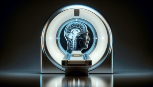
An MRI scan, on the other hand, is a non-invasive medical imaging technique that uses a powerful magnetic field and radio waves to generate detailed images of the internal structures of the body. MRI scans are commonly used to diagnose various medical conditions, including migraines. By understanding the relationship between migraines and MRI scans, we can gain valuable insights into the causes, symptoms, and treatment options for this debilitating condition.
Understanding migraines
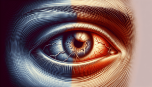
Before delving into how MRI scans can assist in diagnosing migraines, it’s crucial to have a thorough understanding of the condition itself. Migraines are not just ordinary headaches; they are more intense and often come with additional symptoms. Common symptoms of migraines include severe headache pain, nausea, vomiting, sensitivity to light and sound, and visual disturbances.
The exact causes of migraines are still not fully understood, but several contributing factors have been identified. They can be triggered by various factors such as stress, hormonal changes, certain foods or drinks, changes in sleep patterns, or environmental factors. Migraine treatment options can vary depending on the severity and frequency of the episodes, ranging from over-the-counter pain relievers to prescribed medications and lifestyle changes.
MRI scans for migraines
MRI scans are incredibly valuable tools when it comes to diagnosing migraines. They can provide detailed images of the brain structures, allowing healthcare professionals to evaluate if there are any abnormalities or structural changes associated with migraines. By pinpointing these changes, doctors can make more informed decisions regarding treatment plans and management strategies.
An MRI scan can reveal a variety of factors related to migraines. It can show any structural abnormalities in the brain, such as tumors or blood vessel malformations, that may be causing or contributing to migraines. Additionally, MRI scans can detect any signs of inflammation, such as swelling of the blood vessels or abnormal levels of cerebrospinal fluid, which are frequently observed in migraines.
There are different types of MRI scans specifically designed for diagnosing migraines. One such type is a functional MRI (fMRI) scan, which can assess the brain’s functionality during migraine episodes. This scan helps researchers understand the specific areas of the brain that are affected by migraines, leading to a better understanding of the condition and potential treatment options.
While MRI scans are highly valuable in diagnosing migraines, it’s important to note that they do have some limitations. MRI scans cannot directly detect the presence of a migraine during an episode. They are primarily used to rule out other potential causes or identify structural changes associated with migraines. Therefore, a clinical evaluation and medical history assessment are crucial in conjunction with MRI scans for an accurate diagnosis.
Research on MRI and migraines
Over the years, several studies have been conducted to explore the relationship between migraines and brain structure using MRI scans. These studies have provided valuable insights into the effects of migraines on the brain and potential patterns or abnormalities associated with this condition.
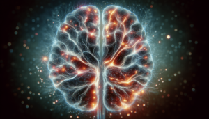
Findings from MRI studies on migraines have shown that individuals who suffer from migraines tend to have alterations in brain structure compared to those without migraines. These alterations can be observed in areas such as the cortex, gray matter, and certain parts of the brain associated with pain modulation and sensory processing.
One such study conducted by researchers in Denmark revealed that migraine sufferers had a thicker cortex in some areas of the brain when compared to non-migraine sufferers. This finding suggests that there may be neuroplastic changes occurring in the brains of individuals with migraines, leading to an increased susceptibility to these debilitating episodes.
Another study conducted at the University of California, San Francisco, demonstrated that individuals with migraines had a higher prevalence of white matter abnormalities, which are linked to changes in brain connectivity. This suggests that migraines may have far-reaching effects on the brain network, potentially leading to the characteristic symptoms experienced during episodes.
Other imaging techniques for migraines
While MRI scans are commonly used to diagnose migraines, there are other imaging techniques available as well. These techniques can be used in conjunction with MRI scans or as alternatives depending on the specific needs and preferences of each patient.
CT scans, or computed tomography scans, are one such alternative imaging technique. CT scans use X-ray technology to generate detailed images of different body structures, including the brain. While they are often used in emergency situations where immediate imaging is required, such as ruling out brain hemorrhages or other acute conditions, they are not as commonly used for diagnosing migraines.
Ultrasound, another imaging technique, utilizes sound waves to create images of the brain and blood vessels. Despite being a non-invasive and relatively low-cost option, ultrasound is not typically used as the primary imaging tool for diagnosing migraines. It may, however, be useful in specific cases where other imaging methods are not feasible or readily available.
PET scans, or positron emission tomography scans, are another option for imaging migraines. PET scans involve the injection of a radioactive tracer that targets specific molecules in the brain. These scans can provide insights into brain metabolism and blood flow, which are helpful in understanding the underlying mechanisms of migraines. However, PET scans are less commonly used due to the availability of less invasive options like MRI scans.
When an MRI is recommended for migraines
While migraines can often be diagnosed based on clinical evaluation and medical history, there are certain situations where an MRI scan may be recommended. These situations typically involve red flags or specific criteria that raise concerns about other potential underlying causes or complications.
One such situation is when considering a diagnosis of “migraine with aura,” where individuals experience specific sensory disturbances before or during their migraines, such as visual disturbances or tingling sensations. In such cases, an MRI scan may be recommended to rule out the possibility of other conditions, such as stroke or brain tumors, which may present with similar symptoms.
Red flags that may prompt an MRI scan include sudden, severe headaches that differ from a person’s usual migraine pattern, headaches associated with neurological deficits (e.g., weakness, numbness, difficulty speaking), or headaches that occur for the first time in older age (over 50). These red flags indicate the need for further investigation to ensure that there are no underlying serious conditions causing the headaches.
Criteria for referral to an MRI scan for migraines may vary depending on the healthcare provider and the specific circumstances. However, it is generally agreed upon that most individuals with migraines can be adequately diagnosed and managed without the need for an MRI scan unless specific concerns arise.
Preparing for an MRI scan for migraines
If you and your healthcare provider have decided that an MRI scan is necessary to further evaluate your migraines, it’s essential to know what to expect and how to prepare for the procedure. MRI scans are painless and non-invasive, but they do require some preparation to ensure accurate and safe imaging.
During an MRI scan, you will need to lie still on a table that will slide into the MRI machine. The machine itself is quite large and may generate loud knocking or buzzing noises during the scan. Earplugs or headphones may be provided to help minimize the noise. The entire process typically takes around 30 to 60 minutes, depending on the specific imaging requirements.
In terms of risks, MRI scans are generally considered safe. However, it’s crucial to inform your healthcare provider if you have any metal implants or devices in your body, as they may interfere with the scan or be affected by the magnetic field. Additionally, if you have a fear of enclosed spaces (claustrophobia) or are unable to lie still for an extended period, let your healthcare provider know in advance so appropriate measures can be taken to ensure your comfort.
To prepare for an MRI scan for migraines, you may be required to remove any metal objects, such as jewelry or hairpins, as they can interfere with the imaging. You may also need to switch into a hospital gown or clothing without metal fasteners. It’s essential to follow any specific instructions given to you by your healthcare provider to ensure a smooth and accurate MRI scan.
Interpreting an MRI scan for migraines
After your MRI scan for migraines, the images will be analyzed and interpreted by a radiologist or neurologist. Reading an MRI scan requires expertise in recognizing normal brain structures as well as abnormalities or structural changes that may be associated with migraines.
The radiologist or neurologist will carefully review the images to identify any anomalies that may contribute to your migraines. They will assess the overall brain structure, comparing it to normal patterns. Areas of interest may include the cortex, gray matter, and regions associated with pain processing and sensory integration.
If any abnormalities or structural changes are identified, your healthcare provider will discuss the findings with you. This discussion may involve additional diagnostic tests, further evaluation, or treatment modifications based on the specific results and your individual situation. Understanding the interpretation of your MRI scan is essential for guiding appropriate treatment and management strategies moving forward.
Alternative methods for diagnosing migraines
While MRI scans are valuable tools in diagnosing migraines, there are alternative methods that healthcare providers may use depending on the circumstances. These methods are particularly useful when MRI scans are unavailable, contraindicated, or when a more cost-effective and accessible approach is desired.
Clinical evaluation plays a vital role in diagnosing migraines. Healthcare providers often rely on their expertise in assessing symptoms, pattern recognition, and medical history to arrive at a diagnosis. They will ask detailed questions about your headaches, including the frequency, intensity, duration, associated symptoms, and any triggers or relieving factors.
Medical history and symptom reporting are also crucial components of diagnosing migraines. Healthcare providers may ask about any family history of migraines, the age of onset, previous treatments, and the success or failure of those treatments. Accurate and detailed reporting of your symptoms can assist your healthcare provider in making an informed diagnosis and developing an appropriate treatment plan.
Keeping a headache diary can be immensely helpful in both diagnosing and managing migraines. A headache diary involves tracking and documenting your headache episodes, including when they occur, the duration, severity, associated symptoms, and any potential triggers or patterns. This information can provide valuable insights into your migraines and assist in evaluating treatment effectiveness or identifying potential changes in your headache pattern over time.
Conclusion
MRI scans play a crucial role in the comprehensive diagnosis and management of migraines. They provide healthcare providers with detailed images of the brain structures, helping identify any abnormalities or structural changes associated with migraines. Through ongoing research, we continue to deepen our understanding of the relationship between migraines and brain structure, which may lead to advancements in treatments and targeted interventions.
While MRI scans are a valuable tool, they are not always necessary for diagnosing migraines. Clinical evaluations, medical history assessments, and symptom reporting are often sufficient for a confident diagnosis. Alternative imaging techniques, such as CT scans, ultrasound, or PET scans, may also be utilized when appropriate or when MRI scans are not readily accessible.
Moving forward, further research is needed to better understand the underlying mechanisms of migraines and how they impact brain structure and function. Advances in imaging technology, such as functional MRI scans or other emerging techniques, may provide even more precise and insightful information about migraines.
If you are suffering from migraines, it’s important to consult with a healthcare professional to discuss your symptoms, receive an accurate diagnosis, and explore the most suitable treatment options. With the help of MRI scans and other diagnostic tools, along with ongoing research and advancements in the field, we can work towards better solutions and improved quality of life for individuals living with migraines.